Imaging Biomarkers
The integration of medical imaging into the clinical Radiation Oncology workflow is rapidly advancing. Computed tomography (CT), as well as a gamut of other imaging modalities — namely, magnetic resonance imaging (MRI) and positron emission tomography (PET) — are now mainstay in Radiation Oncology Departments. Advancement of imaging methods and their application in Radiation Oncology have led to better delineation of targets, such as highly metabolic regions in PET images, the ability to change patient’s treatment plan day-to-day using onboard imaging, and to follow patients’ post-treatment to evaluate treatment response. Our work focuses on incorporating multimodal imaging, such as dynamic contrast enhanced (DCE) CT and intravoxel incoherent motion (IVIM) MRI, to better predict treatment response. Additionally, we are focusing on leveraging advanced image processing techniques, such as deep learning and machine learning, to extract functional information from anatomical images to help guide radiotherapy as well as to improve delineation and tracking of difficult to see targets during image guided interventions. In collaboration with Radiology at UCSF, we are developing lung MR imaging protocols, such as 4DMRI, to extract functional information from free-breathing proton MRI.


Motion Management Systems
Paramount to the implementation of gating or breath-hold motion management in radiotherapy is the detection of respiratory signals using either internal or external probes to track and monitor respiratory motion. Furthermore, to improve the reproducibility of these methods, audiovisual feedback systems previously developed have shown to improve lung tumor position reproducibility and volume consistency. Unfortunately, most clinical systems are sophisticated, complex, and costly, which impedes the widespread use of these systems, especially in locations where staff and resources are limited. Accordingly, our effort is to develop simple-to-use, easy-to-implement low-cost alternatives to commercially available products with the potential to facilitate the translation of respiratory gated techniques to centers that currently do not have access to respiratory motion management systems, namely in lower-middle income countries (LMICs). Our current projects involve developing applications on smartphones with LiDAR capabilities for surface guided radiation therapy (SGRT) and using artificial intelligence to better assist in predicting motion patterns to reduce latency between measuring motion and radiation delivery.
Versions of Smartphone iOS Applications:
iSGRT Static Localization Accuracy - Displacement
X-Displacement

Y-Displacement
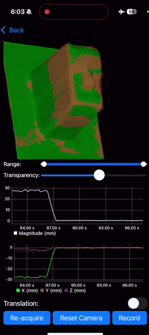
Z-Displacement

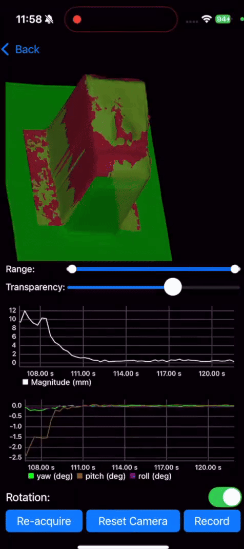
iSGRT Static Localization Accuracy - Rotation
Yaw-Rotation

Pitch-Rotation

Roll-Rotation
Application and schematic illustration of the procedure for acquiring surface imaging.


Quality Assurance Phantoms


With the rapid advancement of technology in Radiation Oncology, there is a growing need for corresponding advancements in quality assurance (QA). QA programs must not only be developed in parallel to ensure the safe and accurate delivery of radiation therapy, but also be designed to ensure that emerging technologies are implemented and utilized to their fullest potential. Given the speed at which novel treatment techniques are translating into clinical practice, rapid prototyping of QA phantoms is becoming desirable—if not essential. Three-dimensional (3D) printing offers a broad range of materials and manufacturing techniques that enable the development of customized QA phantoms tailored to specific clinical and research needs. Our group has access to multiple 3D printing platforms, allowing for rapid design, testing, and iteration of QA phantoms and patient-specific devices for clinical applications. Current research efforts include the testing and refinement of a novel 3D-printed QA phantom, developed in collaboration with Stanford University, to support the implementation of a frameless single-isocenter, multitarget cranial stereotactic radiosurgery (SRS) program. Additional projects focus on the development of devices to evaluate and validate emerging surface-guided radiation therapy imaging systems. We have also previously explored the integration of 3D printing with robotic platforms to create programmable motion models for the evaluation of motion management strategies.